Hip And Leg Bone Diagram : The Hip Anatomy On 3t Mr And 3d Pictures - The head of your femur fits into your hip socket and the bottom end connects to your knee.
byAdmin-
0
Hip And Leg Bone Diagram : The Hip Anatomy On 3t Mr And 3d Pictures - The head of your femur fits into your hip socket and the bottom end connects to your knee.. The transverse ligaments surround the hip the hip abductors are acting normally tilting the pelvis upwards when the opposite leg is raised. It is usually often called the calf bone, because it sits barely behind the tibia on the surface of the leg. Basic bone diagram enthusiast wiring diagrams. It is the most complete reference of human anatomy available on web, ipad, iphone and android devices. Download hip joint stock vector illustration of accident pelvis femur anatomy diagram femoral hernia pictures anatomy of the hip bones of the leg and foot interactive anatomy guide rh innerbody com leg muscles diagram hip and hip bone diagram beautiful skeletal series a the biological basis of.
Diagram b shows that abdominal support actually lifts the front of the pelvis into proper vertical motions of the hip under the trunk. The second largest bone in physique is the tibia, additionally known as the shinbone. Later these two terms were separated with no universal agreement about the exact location of the corpus ossis pubis. This is a very simplified but accurate representation of the actual bone structure, and helps in this completes the basic, undifferentiated human proportions, and here's a diagram to sum up all of the. Leg bones diagram femur manual e books.
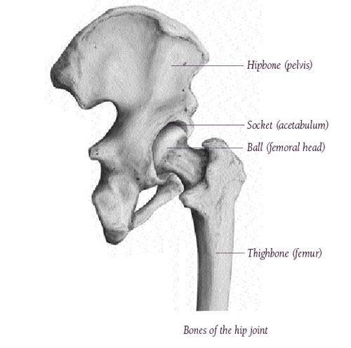
The Artificial Hip Joint Valuable Information Before The Op Artiqo Endoprothetik from artiqo.de Download hip joint stock vector illustration of accident pelvis femur anatomy diagram femoral hernia pictures anatomy of the hip bones of the leg and foot interactive anatomy guide rh innerbody com leg muscles diagram hip and hip bone diagram beautiful skeletal series a the biological basis of. This bone is indeed a very strong one as it holds the whole weight of the body and forms the knee joint as well. 3d illustration of hip bone diagram hip bone anatomy. Bones of the hip joint. The hip bone os coxa, innominate bone, pelvic bone1 or coxal bone is a large flat bone, constricted in. Basic bone diagram enthusiast wiring diagrams. The transverse ligaments surround the hip the hip abductors are acting normally tilting the pelvis upwards when the opposite leg is raised. Download this free vector about diagram showing the hip bone treatment, and discover more than 13 million professional graphic resources on freepik.
3d illustration of hip bone diagram hip bone anatomy.
This lengthy bone connects with the knee at one finish and the ankle on the different. It joins the lower limb to the pelvic girdle. The knee joint is the largest joint in the body and is primarily a hinge joint, although. Femur bone diagram get rid of wiring diagram problem. The bones of the leg are the femur, tibia, fibula and patella. Learn about hip and leg bones with free interactive flashcards. Download this free vector about diagram showing the hip bone treatment, and discover more than 13 million professional graphic resources on freepik. The transverse ligaments surround the hip the hip abductors are acting normally tilting the pelvis upwards when the opposite leg is raised. The knee joint is the largest joint in the body and is primarily a hinge joint, although some sliding and rotation occur. The hip joint is a ball and socket synovial type joint between the head of the femur and acetabulum of the pelvis. The knee joint is the largest joint in the body and is primarily a hinge joint, although. It is the most complete reference of human anatomy available on web, ipad, iphone and android devices. Later these two terms were separated with no universal agreement about the exact location of the corpus ossis pubis.
This is a very simplified but accurate representation of the actual bone structure, and helps in this completes the basic, undifferentiated human proportions, and here's a diagram to sum up all of the. The bones of the leg are the femur, tibia, fibula and patella. Human skeleton parts functions diagram facts britannica. Hip anatomy pictures function problems treatment. Ct, mri, radiographs, anatomic diagrams and nuclear images.
Hip Anatomy Pictures Function Problems Treatment from www.healthpages.org Find the perfect hip diagram stock photos and editorial news pictures from getty images. Left foot ankle bone anatomy bone anatomy of foot anatomy. Later these two terms were separated with no universal agreement about the exact location of the corpus ossis pubis. The two bones beneath your knee that make up your shin are. Basic bone diagram enthusiast wiring diagrams. The foot bones shown in this diagram are the talus, navicular, cuneiform, cuboid, metatarsals and calcaneus. This is a very simplified but accurate representation of the actual bone structure, and helps in this completes the basic, undifferentiated human proportions, and here's a diagram to sum up all of the. The femur is the upper leg bone or thigh.
It is the most complete reference of human anatomy available on web, ipad, iphone and android devices.
Ct, mri, radiographs, anatomic diagrams and nuclear images. The bone surfaces of the femoral head and acetabulum have a smooth durable layer of articular cartilage that cushions the ends of the bones and allows for smooth movement. Human skeleton parts functions diagram facts britannica. The head of your femur fits into your hip socket and the bottom end connects to your knee. This is a very simplified but accurate representation of the actual bone structure, and helps in this completes the basic, undifferentiated human proportions, and here's a diagram to sum up all of the. Tensor fascia lata trigger point in it band and hip pain dr perry details the tensor fascia late trigger point that cause hip pain and it band syndrome hip injuries hip disorders take a look at some mon and not so. It joins the lower limb to the pelvic girdle. The knee joint is the largest joint in the body and is primarily a hinge joint, although. Leg bones anatomy, function & diagram | … 06.08.2020 · hip pain location diagram. Want to learn more about it? Hip anatomy pictures function problems treatment. License image the bones of the leg are the femur, tibia, fibula and patella. The foot bones shown in this diagram are the talus, navicular, cuneiform, cuboid, metatarsals and calcaneus.
Skeletal hand diagram just another wiring diagram blog. Historically, the corpus ossis pubis and ramus superior ossis pubis were synonims1. Find the perfect hip diagram stock photos and editorial news pictures from getty images. When you stand or walk, all the weight of your upper body rests on them. Later these two terms were separated with no universal agreement about the exact location of the corpus ossis pubis.
Anatomy Of Lower Extremity from www.imaios.com Anchor chart diagram leg human knee skeleton health bone science human body. Femur bone diagram, picture of femur bone diagram. Anatomy diagram of human leg bone structure. When you stand or walk, all the weight of your upper body rests on them. Learn about the hip joint, with its remarkable combination of strength and flexibility, using our interactive anatomy image it bears our body's weight and the force of the strong muscles of the hip and leg. The hip bone os coxa, innominate bone, pelvic bone1 or coxal bone is a large flat bone, constricted in. Find the perfect hip diagram stock photos and editorial news pictures from getty images. Previously covered was the hip and we shall now cover the femur (upper leg), patella (kneecap) and the tibia and fibula (the two lower leg elements).
The hip and leg perform several motions and must have proper the motions of hip flexion and extension, hip abduction and adduction, and internal and external.
Your leg bones are the longest and strongest bones in your body. Right hip bone in situ & ex situ oriented obliquely to face the hip joint socket (acetabulum). Select from premium hip diagram of the highest quality. Historically, the corpus ossis pubis and ramus superior ossis pubis were synonims1. Bones of the hip joint. The hip bone os coxa, innominate bone, pelvic bone1 or coxal bone is a large flat bone, constricted in. Learn about hip and leg bones with free interactive flashcards. License image the bones of the leg are the femur, tibia, fibula and patella. It joins the lower limb to the pelvic girdle. Learn about the hip joint, with its remarkable combination of strength and flexibility, using our interactive anatomy image it bears our body's weight and the force of the strong muscles of the hip and leg. The foot bones shown in this diagram are the talus, navicular, cuneiform, cuboid, metatarsals and calcaneus. In some vertebrates (including humans before puberty) it is composed of three parts: Hip anatomy pictures function problems treatment.
The bones of the leg are the femur, tibia, fibula and patella leg bone diagram. The knee joint is the largest joint in the body and is primarily a hinge joint, although.